The phrase “larger than life” many times throughout history has been applied to describe the impact of scientific discoveries and revolutionary technologies. At the University of Notre Dame, precision instruments and state-of-the-art facilities, such as the electron microscopy core within the Integrated Imaging Facility, are beginning to fill in gaps and reveal details that are propelling science beyond what is known.
Sharon Stack, Ann F. Dunne and Elizabeth Riley Director of the Harper Cancer Research Institute and Kleiderer-Pezold Professor of Chemistry and Biochemistry, uses the University’s advanced electron microscopy core (EM) to view the intricate surface of cancer cells. One such study examined the correlation of obesity and the role that it plays on the progression of ovarian cancer whereby the researchers discovered a striking change in peritoneal ultrastructure, with a substantial increase in mesothelial microvilli content on the surface of cells that could not be visualized by an optical microscope alone
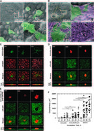
“Our research evaluating molecular mechanisms of ovarian cancer metastasis has benefitted significantly from use of the EM Core. The facility has enabled us to open new directions in our research program based on the level of ultra-structural detail we are able to visualize,” Stack said.
Sergei Rouvimov, Transmission Electron Microscopy (TEM) director at Notre Dame is dedicated in solving problems presented by previous limitations and often uses a combination of techniques during the process of evaluation. Rouvimov explains that the field of electron microscopy is
“delicate and can offer potential solutions to researchers that are far beyond what is known from previous methods.”
One such researcher using the facility to further examine properties and structural changes within nanotube and nanoribbon development, which have a wide range of uses such as optoelectronic and electronic devices including solar cells, photodetectors, sensors, field-effect transistors (FETs), and logic circuits, is Alan Seabaugh. Seabaugh, Director of NDnano and Frank M. Freimann Chair Professor of Electrical Engineering, said,
“The single most informative characterization we make is with transmission electron microscopy. Without this imaging we would be unable to see if our processes have worked as designed.”

The University of Notre Dame has long-valued the impact that these sophisticated microscopes can have when coupled with world-class researchers and students. And, earlier this year, the world recognized them as well, when three scientists from Switzerland, USA, and United Kingdom, were announced as winners of a Nobel Prize in Chemistry “for developing cryo-electron microscopy for the high-resolution structure determination of biomolecules in solution.” It was the legacy of these three scientists and their impact on the field that will continue to propel and inspire researchers in the field of electron microscopy.
The Notre Dame Integrated Imaging Facility provides an integrated suite of sophisticated microscopes and imaging stations, including electron microscopy, biological imaging (in vivo and optical microscopy), and histology, that enable expert users to attack the most complex modern research problems and, equally important, resident professional staff, including technicians and research specialists, to guide the non-expert users. The facility is open to the Notre Dame research community, external academic institutions, and industry. Additionally, it is an approved Indiana Clinical and Translational Sciences Institute (CTSI) core facility, with services available to all CTSI partners. Learn more at imaging.nd.edu.
Originally published by at imaging.nd.edu on December 05, 2017.